


Osteoarthritis of the shoulderIt's chronic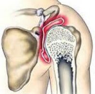 a disease in which the articular cartilage tissue is destroyed and thinned, pathological changes in the soft tissues occur, and bone growths form in the joint area. It is manifested by pain and crunch in the affected area. In the later stages, the range of motion decreases. The pathology is chronic and gradually progresses. The diagnosis is made taking into account the clinical picture and radiological signs. Treatment is usually conservative: physiotherapy, anti-inflammatory drugs, chondroprotectors, exercise therapy. When the joint is destroyed, arthroplasty is performed.
General information
Osteoarthritis of the shoulder joint is a chronic disease in which, as a result of degenerative-dystrophic processes, cartilage and other tissues of the joint are gradually destroyed. Usually osteoarthritis affects people over the age of 45, but in some cases (after injury, inflammation) the disease can develop at a younger age. The pathology occurs equally often in women and men, it is more often noticed in athletes and in people who do hard physical work.
Reasons
The starting point for changes in arthrosis of the shoulder joint can be a normal process of tissue aging and damage or disruption of cartilage structure as a result of mechanical influences and various pathological processes. Primary arthrosis is usually detected in the elderly, secondary (developed against the background of other diseases) can occur at any age. The main reasons are taken into account:
- Developmental defects.Pathology can be detected in the underdevelopment of the head of the humerus or glenoid cavity, capomelia of the shoulder and other anomalies of the upper extremity.
- Traumatic injury.Osteoarthritis of traumatic etiology most often occurs after intra-articular fractures. A possible cause of the disease can be a dislocation of the shoulder, especially the usual one. Rarely, severe bruising acts as a provocative injury.
- Inflammatory processes.The disease can be diagnosed with long-term shoulder-scapular periarthritis, previously suffered nonspecific purulent arthritis and specific joint arthritis (with tuberculosis, syphilis and some other diseases).
Risk factors
Osteoarthritis is a polyetiological disease. There is a wide group of factors that increase the likelihood of this pathology:
- Hereditary predisposition.Many patients have close relatives who also suffer from arthrosis, including those of other localizations (gonarthrosis, coxarthrosis, arthrosis of the ankle joint).
- Joint strain.It can occur in volleyball players, tennis players, basketball players, sports equipment throwers, as well as in people whose profession involves a constant heavy load on the hands (hammers, loaders).
- Other pathologies. Osteoarthritis is more common in patients suffering from autoimmune (rheumatoid arthritis), some endocrine diseases and metabolic disorders, systemic connective tissue insufficiency and excessive joint mobility.
The likelihood of developing the disease increases dramatically with age. Frequent hypothermia and unfavorable environmental conditions have a certain negative impact.
Pathogenesis
The main reason for the development of arthrosis of the shoulder joint is a change in the structure of the articular cartilage. Cartilage loses its smoothness and elasticity, sliding of joint surfaces during movement becomes difficult. Microtraumas occur which lead to further deterioration of the condition of the cartilage tissue. Small pieces of cartilage separate from the surface, forming free-lying articular bodies, which also injure the inner surface of the joint.
Over time, the capsule and synovium thicken, and areas of fibrous degeneration appear in them. Due to the thinning and reduction of elasticity, the cartilage ceases to provide the necessary shock absorption, therefore the load on the lower bone increases. Bone deforms and grows along the edge. The normal configuration of the joint is disturbed, there are restrictions on movement.
Classification
In traumatology and orthopedics, a three-stage systematization is usually used, which reflects the severity of pathological changes and symptoms of arthrosis of the shoulder joint. This approach allows you to choose the optimal medical tactics, taking into account the difficulty of the process. The following phases are distinguished:
- The first- no gross changes in cartilage tissue. The composition of the synovial fluid is changed, the nutrition of the cartilage is damaged. Cartilage does not tolerate stress, so from time to time joint pain (arthralgia) occurs.
- Others- cartilage tissue begins to thin, its structure changes, the surface loses smoothness, cysts and areas of calcification appear in the depth of the cartilage. The lower bone is slightly deformed, bone growths appear on the edges of the articular platform. The pain becomes permanent.
- Third- marked thinning and disruption of the cartilage structure with large areas of destruction. The articulated platform is deformed. Limitation of range of motion, weakness of the ligament apparatus and atrophy of the periarticular muscles were detected.
Symptoms
In the early stages, patients with osteoarthritis worry about discomfort or minor pain in the shoulder joint during exertion and certain body positions. Crushing may occur during movement. The joint is not externally altered, there is no edema. Then the intensity of the pain increases, arthralgias become common, permanent, they appear not only during exercise, but also at rest, including at night. Characteristic characteristics of the pain syndrome:
- Many patients notice the dependence of pain syndrome on weather conditions.
- Along with excruciating pain, over time there is sharp pain during physical exertion.
- The pain may appear only in the shoulder joint, radiate to the elbow joint, or spread throughout the arm. Possible back and neck pain on the affected side.
After a while, patients begin to notice a noticeable morning stiffness in the joint. The range of motion decreases. Slight swelling of the soft tissues is possible after exercise or hypothermia. As osteoarthritis progresses, movements become more limited, contractures develop, and limb function is severely impaired.
Diagnosis
The diagnosis is made by an orthopedic surgeon, taking into account the characteristic clinical and radiological signs of arthrosis of the shoulder joint. If you suspect secondary osteoarthritis, consult a surgeon, endocrinologist. At first the joint does not change, later it sometimes deforms or enlarges. Palpation reveals pain. Restriction of movement can be detected. To confirm osteoarthritis, the following are recommended:
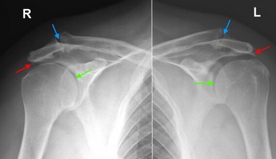
- Radiography of the shoulder joint.Dystrophic changes and marginal bone growths (osteophytes) were found, in later stages narrowing of the joint space, deformations and changes in the structure of the lower bone were determined. The joint cavity may take a wedge shape, osteosclerotic changes and cystic formations are visible in the bone.
- Tomographic examination.In suspicious cases, especially in the initial stages of the disease, CT of the shoulder joint is performed in order to obtain additional data on the condition of the bones and cartilage. If it is necessary to assess the condition of the soft tissues, magnetic resonance imaging is performed.
Differential diagnosis
The differential diagnosis of arthrosis is made in gout, psoriasis, rheumatoid and reactive arthritis, as well as pyrophosphate arthropathy. In arthritis, a blood test shows signs of inflammation; changes on the radiograph are not very pronounced, osteophytes are absent, there are no signs of deformation of the articular surfaces.
In psoriatic arthritis, along with joint manifestations, skin rashes can often be found. In rheumatoid arthritis, a positive rheumatoid factor is determined. With pyrophosphate arthropathy and gouty arthritis, a biochemical blood test reveals corresponding changes (increase in uric acid salt levels, etc. ).
Treatment of shoulder arthrosis
Patients are under the supervision of an orthopedic surgeon. It is necessary to limit the load on the arm, excluding sudden movements, lifting and carrying weights for a longer time. At the same time, it should be borne in mind that inaction negatively affects the diseased joint. To keep the muscles in a normal state, as well as to restore the shoulder joint, you must regularly perform an exercise complex recommended by a doctor.
Conservative treatment
One of the most urgent tasks in osteoarthritis is to fight pain. To eliminate pain and reduce inflammation, the following are prescribed:
- Drugs of general action.NSAIDs are prescribed in tablets during exacerbations. Uncontrolled use can irritate the stomach wall, adversely affect the condition of the liver and metabolism in cartilage tissue, so they are taken only as directed by a doctor.
- Local remedies.NSAIDs are usually used in the form of gels and ointments. Self-administration is possible if symptoms appear or intensify. Less often, local hormonal preparations are indicated, which should be used in accordance with the doctor's recommendations.
- Hormones for intra-articular administration.In the case of severe pain syndrome, which cannot be eliminated by other methods, intra-articular administration of drugs (triamcinolone, hydrocortisone, etc. ) is performed. Blockades are performed no more than 4 times a year.
To restore and strengthen cartilage in the 1st and 2nd stage of arthrosis, agents from the group of chondroprotectors are used - drugs that contain hyaluronic acid, chondroitin sulfate and glucosamine. The courses of treatment are long (from 6 months to a year or more), the effect becomes noticeable after 3 or more months.
Physiotherapy treatment
Massage, physiotherapy exercises and physiotherapy techniques are actively used for arthrosis of the shoulder joint. During the period of remission, patients are referred to spa treatment. Apply:
- mud and paraffin therapy;
- medicinal baths;
- magnetotherapy and infrared laser therapy;
- ultrasound.
Surgery
In stage 3 of the disease, with significant destruction of cartilage, limitation of mobility and disability, joint replacement is performed. The recommendation for the operation is given taking into account the age of the patient, the level of his activity, the presence of severe chronic diseases. The use of modern ceramic, plastic and metal endoprostheses allows you to completely restore joint function. The lifespan of the prosthesis is 15 or more years.
Forecast
Osteoarthritis is a long-term, gradually progressive disease. It cannot be completely cured, however, it is possible to significantly slow down the development of pathological changes in the joint, preserve working ability and a high quality of life. To achieve maximum effect, the patient must be serious about his illness and his willingness to follow the doctor’s recommendations, even during the remission period.
Prophylaxis
Preventive measures include reducing injuries in the household, respecting safety at work, removing excessive strain on the shoulder joint when performing professional duties and playing sports. It is necessary to timely diagnose and treat pathologies that can cause the development of arthritic changes.